- OPG
- CBCT
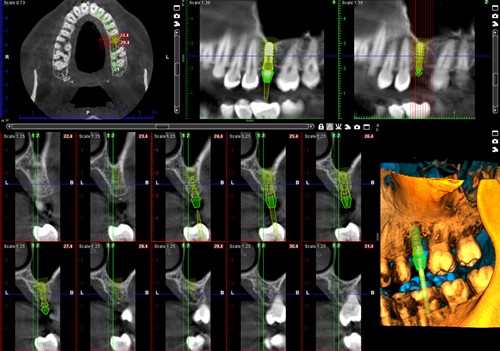
- 3D Implant Planning

Radio diagnosis & Reporting

Home / Radio diagnosis & Reporting

Our team consist of dental professionals with proven track record of success and experience in radiological imaging and patient care. They are highly skilled in interpreting images to diagnose patient's health conditions.

Our services

  • 2D imaging- OPG, TMJ Double Lateral & PA, Hand wrist, Lateral Cephalogram and other skull views
    An OPG is a panoramic X-ray of the upper and lower jaws, including the teeth. The OPG unit is specifically designed to rotate around the patient’s head during the scan and the scan is over in less than a minute.
  • 3D imaging- Cone-beam computed tomography (CBCT) is a radiographic imaging method that allows accurate, three-dimensional (3D) imaging. We have Planmeca ProFace which is a unique way to produce a realistic 3D face photo and a CBCT image with a single scan. A 3D face photo can also be created separately without exposing the patient to any radiation. 3D imaging is generally used for the assessment of maxillofacial and TMJ pathology, implant planning, surgical evaluation of impacted teeth, palatal cleft and facial deformities, air-way analysis and endodontic assessment.

How to approach us

Send the patient directly to the center or mail us the CBCT scan in DICOM format.

How do we respond

48 hours is the turnaround time we keep to generate a comprehensive report that is mailed to your email account in pdf format.

Work Flow

1

Starting Point

Send the patient directly to our center / Mail the CBCT Scan in DICOM format via we-transfer to ddscochin@gmail.com / Courier the data in a portable storage device (CD/Pendrive) to our address.
2

Assessment

Assessment of the data by our Radiologists.
3

Completion

Within 48 hours the report is mailed to the concerned account.

FAQ

2D imaging- OPG, TMJ Double Lateral & PA, Hand wrist, Lateral Cephalogram and other skull views... 3D imaging- CBCT, Facial scan

Mail the CBCT scan in DICOM format via we transfer to ddscochin@gmail.com or courier CD to our center.
Note: Make sure you are sending only the raw DICOM data and not the processed images with the viewer.

8 x 8 - both maxilla & mandible
8 x 5 – single arch (either maxilla or mandible).
5 x 5- single tooth.
15 x 8 - for visualizing zygoma.

Yes, Within 48 hours.

Copyright © 2021-2023. Digital Dental Solutions, Kochi : ddskochi.com | A Dentaura Design